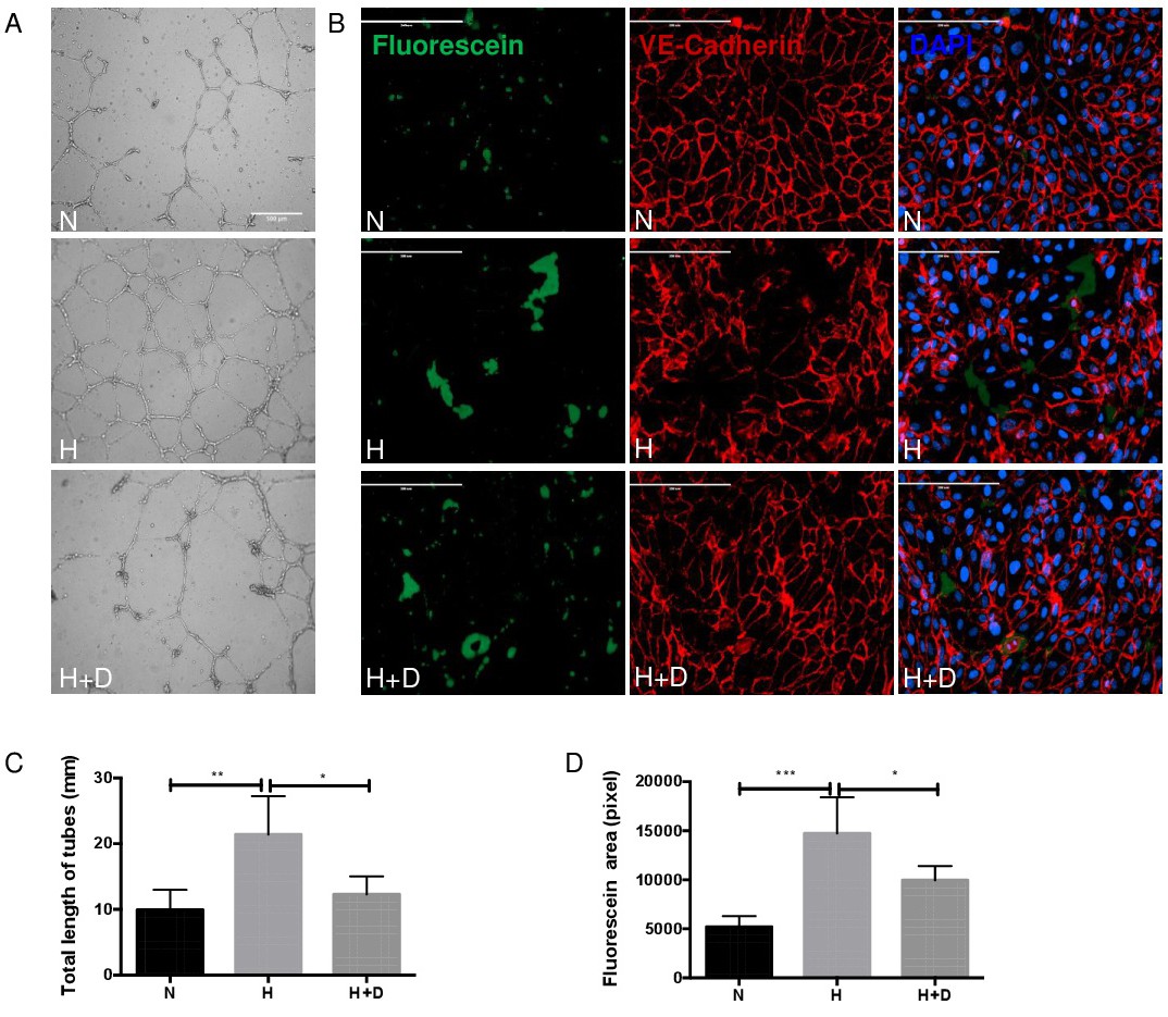
Fig. 2. Conditioned medium from hypoxic MIO-M1 cells promotes angiogenesis and vessel leakage. (A-B) Conditioned media from MIO-M1 cells exposed to 1% O2 hypoxia 48 hours (H) stimulated HRMECs tube formation compared to 20% O2 normoxic condition (N) (n=12 per group). The total length of tubes decreased by pretreatment with digoxin (H+D). Images was performed with Leica DMI6000 microscope (x5). Scale bar, 500 μm. (C-D) The area of FITC-positive intercellular spots increased conditioned medium from hypoxic MIO-M1 cells and decreased by pretreatment with digoxin (n=3 per group). Scale bar, 200 μm. Data represents means ± SD of relative values vs control from 3 independent experiments. *P<0.05; **P<0.01; ***P<0.001; ****P<0.0001, statistical analysis was performed with one-way ANOVA with Dunn's test for multiple comparisons.